
Product
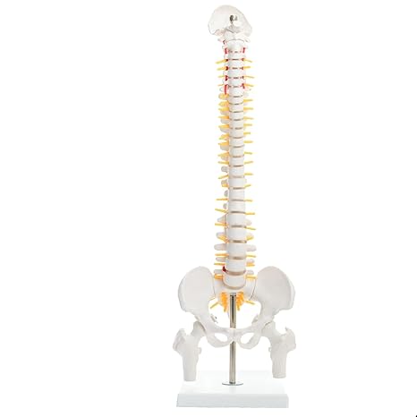
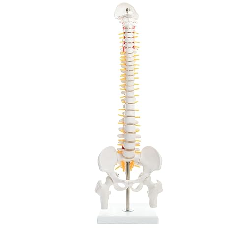
Human Mini Spine Model | Spinal & Vertebral Column Anatomy
₦280,000.00
Human Mini Spine Model is a detailed 45 cm anatomical model of the human vertebral column designed for anatomy, physiology, physiotherapy, medical education, and patient demonstrations. It features the cervical, thoracic, and lumbar vertebrae, sacrum, coccyx, complete pelvis, femur heads, spinal nerves, intervertebral discs, and a representation of a prolapsed L3-L4 disc. Made from durable PVC and mounted on a stable stand, it is suitable for medical schools, clinics, hospitals, physiotherapy centres, laboratories, classrooms, and healthcare offices.
Add to cart
Buy Now
Human Mini Spine Model (Spinal/Vertebral Column Model) provides a three-dimensional representation of the human spine and its relationship with the pelvis and upper femoral structures.
Standing approximately 45 cm tall, this anatomical spine model allows students, healthcare professionals, therapists, and patients to visualize the major sections of the vertebral column and understand how the spine connects with the pelvis.
The model includes the cervical, thoracic, and lumbar vertebrae, as well as the sacrum, coccyx, complete pelvis, femur heads, spinal nerves, and intervertebral discs.
A particularly useful teaching feature is the representation of spinal nerve branches and a prolapsed L3-L4 intervertebral disc, providing a visual reference for explaining basic spinal anatomy and selected spine-related conditions.
The model is mounted on a stand, making it convenient for display on desks, laboratory benches, clinic tables, classroom demonstration areas, and medical offices.
Key Features
- 45 cm Tall Spine Model: Compact but detailed anatomical representation of the human spine.
- Complete Vertebral Column: Includes cervical, thoracic, and lumbar vertebrae.
- Sacrum & Coccyx: Demonstrates the lower sections of the vertebral column.
- Complete Pelvis: Helps students understand the relationship between the spine and pelvic structures.
- Detachable Femur Heads: Allows closer examination of the pelvic and hip anatomy.
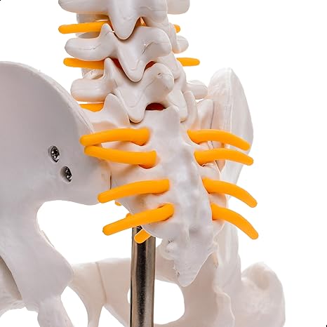
- Spinal Nerves: Includes a representation of spinal nerve branches.
- Intervertebral Discs: Shows the discs located between the vertebrae.
- L3-L4 Prolapsed Disc Representation: Demonstrates a selected spinal disc pathology for educational purposes.
- Anatomically Detailed: Designed to provide a clear visual representation of spinal structures.
- High-Quality PVC: Made from durable PVC plastic for repeated educational use.
- Ivory-Coloured Vertebrae: Contrasting anatomical elements make structures easier to distinguish.
- Mounted on Stand: Provides convenient upright display and classroom demonstration.
- Portable Design: Suitable for offices, classrooms, laboratories, clinics, and therapy centres.
- Educational & Demonstration Tool: Useful for anatomy, physiology, physiotherapy, and patient education.
Anatomical Structures Demonstrated
The Human Mini Spine Model provides a visual representation of several important spinal and pelvic structures, including:
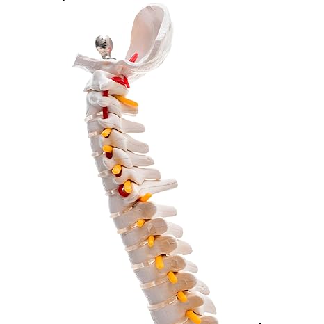
Cervical Vertebrae
The cervical region represents the upper portion of the vertebral column and can be used to introduce students to the anatomy of the neck and upper spine.
Thoracic Vertebrae
The thoracic section demonstrates the middle portion of the spinal column and its relationship to the surrounding trunk anatomy.
Lumbar Vertebrae
The lumbar section provides a visual reference for the lower back region and is particularly useful when teaching basic lumbar spine anatomy.
Sacrum
The sacrum demonstrates the connection between the lower spine and the pelvis.
Coccyx
The coccyx, or tailbone, is included as part of the lower vertebral column.
Complete Pelvis
The complete pelvis helps learners understand the anatomical relationship between the vertebral column and pelvic structures.
Femur Heads
The femur heads are detachable, allowing students and educators to examine the relationship between the pelvis and proximal femoral structures.
Intervertebral Discs
The model shows intervertebral discs positioned between vertebral segments, providing a visual aid for explaining spinal movement and disc anatomy.
Spinal Nerve Branches
Representations of spinal nerve branches are included to help explain the relationship between the vertebral column and associated neural structures.
L3-L4 Prolapsed Disc Demonstration
One of the useful educational features of this model is the representation of a prolapsed L3-L4 intervertebral disc.
This allows educators to introduce students to the anatomical concept of disc prolapse and demonstrate how changes involving an intervertebral disc can be discussed in relation to surrounding spinal structures.
The representation should be regarded as an educational anatomical illustration, not as a tool for diagnosing an actual patient’s spinal condition.
Educational Benefits
1. Makes Spine Anatomy Easier to Understand
A three-dimensional model allows students to view the vertebral column from different angles rather than relying solely on textbook diagrams.
2. Demonstrates the Relationship Between Spine and Pelvis
The inclusion of the complete pelvis and femur heads helps students visualize the connection between the vertebral column, pelvic region, and hip structures.
3. Supports Physiotherapy Education
Physiotherapy students and educators can use the model when introducing basic spinal anatomy and selected musculoskeletal concepts.
4. Useful for Anatomy & Physiology Classes
The model provides a practical visual aid for explaining the different sections of the vertebral column.
5. Supports Patient Demonstrations
Healthcare professionals can use the model to visually explain basic spinal structures to patients during consultations or educational sessions.
6. Demonstrates Spinal Nerves
The spinal nerve representations provide an additional visual reference when discussing the relationship between vertebrae and neural structures.
7. Helps Explain Intervertebral Discs
The model allows learners to identify the location of intervertebral discs and understand their position between adjacent vertebrae.
Common Applications
The Human Mini Spine Model can be used in:
- Medical schools
- Nursing schools
- Physiotherapy schools
- Chiropractic education
- Anatomy laboratories
- Hospitals
- Clinics
- Physiotherapy centres
- Rehabilitation centres
- Medical training institutions
- Healthcare offices
- Patient education
- Biology and physiology classrooms
- Medical exhibitions
- Healthcare training programmes
Suitable for Physiotherapy & Rehabilitation Teaching
This spinal anatomical model can be particularly useful for teaching introductory concepts involving:
- Vertebral column anatomy
- Cervical spine anatomy
- Thoracic spine anatomy
- Lumbar spine anatomy
- Sacral anatomy
- Coccyx anatomy
- Pelvic anatomy
- Intervertebral discs
- Spinal nerve anatomy
- Lumbar disc prolapse
- Spine and pelvis relationship
- Basic musculoskeletal anatomy
For clinical decision-making, diagnosis, treatment planning, or surgical procedures, qualified healthcare professionals should rely on appropriate clinical assessment, imaging, and other relevant medical information rather than the model alone.
Durable PVC Construction
The model is made from high-quality PVC plastic, providing a durable construction suitable for repeated classroom and professional demonstrations.
The ivory-coloured vertebrae contrast with other anatomical elements, helping students distinguish different structures more easily.
Its compact size and mounted stand also make it convenient for keeping on a desk, clinic table, laboratory bench, or classroom demonstration area.
Detachable Femur Heads
The femur heads are detachable, providing additional flexibility when demonstrating the relationship between the pelvis and femoral structures.
This feature can be particularly useful during anatomy lessons where students need to examine the pelvic and hip region more closely.
Specifications
| Specification | Details |
|---|---|
| Product Name | Human Mini Spine Model |
| Alternative Name | Spinal / Vertebral Column Anatomical Model |
| Product Type | Human Spine & Pelvis Anatomical Model |
| Height | Approximately 45 cm |
| Main Anatomy | Human Vertebral Column |
| Cervical Vertebrae | Included |
| Thoracic Vertebrae | Included |
| Lumbar Vertebrae | Included |
| Sacrum | Included |
| Coccyx | Included |
| Pelvis | Complete pelvis included |
| Femur Heads | Detachable |
| Intervertebral Discs | Included |
| Spinal Nerves | Spinal nerve branches represented |
| Disc Pathology | Prolapsed L3-L4 disc representation |
| Material | High-quality PVC plastic |
| Colour | Ivory-coloured vertebrae with contrasting anatomical elements |
| Mounting | Mounted on stand |
| Construction | 3D anatomical model |
| Primary Use | Anatomy, physiology & healthcare education |
| Suitable Settings | Schools, Clinics, Hospitals, Laboratories & Therapy Centres |
How to Use the Spine Model
The model is intended primarily for educational and demonstration purposes.
A teacher, lecturer, physiotherapist, or healthcare professional can:
- Place the model securely on a stable surface.
- Introduce the major regions of the vertebral column.
- Identify the cervical, thoracic, and lumbar vertebrae.
- Demonstrate the sacrum and coccyx.
- Explain the relationship between the spine and complete pelvis.
- Detach the femur heads when demonstrating pelvic and hip anatomy.
- Identify the intervertebral discs.
- Demonstrate the spinal nerve branches represented on the model.
- Use the L3-L4 prolapsed disc representation to introduce disc pathology.
- Relate the model to textbooks, diagrams, lectures, clinical education, or patient demonstrations.
Why Choose This Human Mini Spine Model?
The Human Mini Spine Model combines major spinal anatomy, pelvic structures, intervertebral discs, spinal nerve representations, and a selected disc pathology in one compact teaching model.
Its 45 cm height, durable PVC construction, detachable femur heads, mounted stand, and contrasting anatomical structures make it practical for repeated demonstrations.
It can be an excellent addition to:
- Medical anatomy laboratories
- Physiotherapy departments
- Nursing schools
- Medical training institutions
- Hospitals and clinics
- Rehabilitation centres
- Healthcare offices
- Patient education programmes
Clinical & Research Applications
Anatomical spine models can also serve as visual aids when discussing spinal conditions, treatment concepts, biomechanics, and procedures.
This model may be useful for introductory demonstrations related to spinal anatomy and can provide a physical reference during discussions involving the vertebral column and pelvis.
For clinical diagnosis, surgical planning, implant selection, biomechanical testing, or medical research, this educational model should be used only as a supplementary visual aid alongside appropriate professional resources, clinical data, imaging, and manufacturer documentation.
Care & Storage
- Place the model on a stable surface during demonstrations.
- Handle detachable femur heads carefully.
- Avoid dropping or applying excessive force to anatomical components.
- Keep away from excessive heat and prolonged direct sunlight.
- Clean using a method appropriate for PVC.
- Avoid harsh chemicals unless confirmed safe by the manufacturer.
- Store in a clean, dry location when not in use.
- Keep the model securely mounted on its stand during display.
- Use the model primarily for educational and demonstration purposes.
Frequently Asked Questions
1. What is the Human Mini Spine Model used for?
It is used as an anatomical teaching aid for spine anatomy, physiology, physiotherapy, rehabilitation education, classroom demonstrations, and patient education.
2. How tall is the spine model?
The model is approximately 45 cm tall.
3. Which parts of the spine are included?
It includes the cervical, thoracic, and lumbar vertebrae, sacrum, and coccyx.
4. Does the model include the pelvis?
Yes. The model includes a complete pelvis to demonstrate the relationship between the spine and pelvic structures.
5. Are the femur heads detachable?
Yes. The femur heads are detachable, allowing additional examination of the pelvic and hip anatomy.
6. Does the model show spinal nerves?
Yes. The model includes a representation of spinal nerve branches.
7. Does it show intervertebral discs?
Yes. Intervertebral discs are represented between the vertebral structures.
8. Does the model demonstrate a prolapsed disc?
Yes. It includes a representation of a prolapsed L3-L4 intervertebral disc for educational demonstration.
9. What material is the spine model made from?
The supplied specifications state that it is made from high-quality PVC plastic.
10. Does the spine model come with a stand?
Yes. The model is mounted on a stand for convenient display and teaching.
11. Can physiotherapy students use this model?
Yes. It is suitable for physiotherapy and rehabilitation education, particularly when teaching basic spinal, pelvic, disc, and musculoskeletal anatomy.
12. Can the model be used to diagnose a slipped or prolapsed disc?
No. It is an educational anatomical model. Actual spinal conditions require appropriate assessment by a qualified healthcare professional and, where necessary, diagnostic imaging.
You may also like…


Related products