
Product
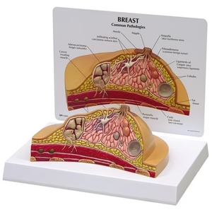
Full size Breast Cross Section ModelBreast Cross Section Model
₦150,000.00
Full Size Breast Cross Section Model with Pathologies is a detailed anatomical teaching model designed to demonstrate the internal structures of the human breast and common breast pathologies. The model illustrates conditions including adenocarcinoma, cysts, fibroadenoma, and infiltrating scirrhus carcinoma, while also showing important anatomical structures such as suspensory ligaments, fat tissue, lymph nodes, muscles, and ribs. It is ideal for medical schools, nursing education, breast health teaching, anatomy classes, pathology training, and healthcare professional demonstrations.
Add to cart
Buy Now
Full Size Breast Cross Section Model provides a three-dimensional representation of breast anatomy combined with visual examples of several commonly studied breast pathologies.
Designed as an educational tool, the model allows students and healthcare professionals to examine the relationship between normal breast structures and selected pathological changes.
The cross-sectional design exposes internal structures that are difficult to visualize using a conventional external breast model. It can therefore be used to support teaching about breast anatomy, breast pathology, lymphatic structures, and the location and appearance of selected lesions.
The model depicts:
- Adenocarcinoma
- Cysts
- Fibroadenoma
- Infiltrating scirrhus carcinoma
- Suspensory ligaments
- Fat tissue
- Lymph nodes
- Muscles
- Ribs
This makes it a practical visual aid for institutions teaching anatomy, pathology, nursing, medicine, breast health, and related healthcare subjects.
Key Features
- Full-Size Breast Cross Section: Provides a detailed sectional representation of breast anatomy.
- Pathology Demonstration: Displays several common breast pathologies for educational study.
- Adenocarcinoma Representation: Includes an anatomical representation of adenocarcinoma.
- Cyst Representation: Shows breast cysts as part of the pathology demonstration.
- Fibroadenoma Representation: Includes fibroadenoma for comparative pathology education.
- Infiltrating Scirrhus Carcinoma: Demonstrates this pathology within the breast cross section.
- Suspensory Ligaments: Shows supporting connective structures of the breast.
- Fat Tissue: Demonstrates the distribution of fatty tissue within the breast.
- Lymph Nodes: Illustrates lymphatic structures relevant to breast anatomy education.
- Muscles & Ribs: Shows surrounding anatomical structures for better spatial understanding.
- Three-Dimensional Design: Allows learners to study breast structures from a physical model.
- Educational Teaching Aid: Suitable for classroom lectures, laboratory demonstrations, and healthcare training.
- Professional Anatomy Model: Useful for medicine, nursing, pathology, and breast-health education.
Breast Anatomy Demonstration
The model provides a visual representation of the internal structures of the breast.
Students can use it to identify and discuss:
Suspensory Ligaments
The model shows the suspensory ligaments of the breast, helping students understand the supporting structures associated with breast anatomy.
Fat Tissue
The model illustrates fat tissue within the breast, providing a visual reference for understanding the composition and internal organization of breast tissue.
Lymph Nodes
The inclusion of lymph nodes helps educators introduce the relationship between breast anatomy and the lymphatic system.
Muscles
The model shows relevant muscular structures surrounding the breast, providing additional anatomical context.
Ribs
The ribs shown in the cross section help learners understand the position of the breast relative to the underlying thoracic structures.
Breast Pathologies Demonstrated
One of the major advantages of this model is its ability to demonstrate selected breast pathologies alongside normal anatomical structures.
1. Adenocarcinoma
The model includes an anatomical representation of adenocarcinoma, allowing educators to introduce students to malignant breast pathology.
2. Cysts
Breast cysts are represented within the model, providing a visual aid for explaining cystic breast lesions.
3. Fibroadenoma
The model demonstrates fibroadenoma, allowing students to compare this commonly studied benign breast lesion with other breast abnormalities.
4. Infiltrating Scirrhus Carcinoma
The model also depicts infiltrating scirrhus carcinoma, providing an additional example for pathology education.
These representations are intended for educational demonstration and should not be used to diagnose a real patient or determine whether a patient’s breast lesion is benign or malignant.
Educational Benefits
1. Makes Breast Anatomy Easier to Understand
The three-dimensional cross-section allows students to visualize structures that may be difficult to understand from textbook illustrations alone.
2. Supports Pathology Education
The model provides a physical reference for studying selected breast pathologies and comparing their locations and appearances.
3. Useful for Medical Students
Medical students can use the model as a supplementary teaching aid during anatomy and pathology lessons.
4. Supports Nursing Education
Nursing instructors can use the model when teaching breast anatomy, breast health, patient education, and related pathology concepts.
5. Helps Explain Lymphatic Anatomy
The inclusion of lymph nodes allows instructors to introduce the anatomical relationship between breast tissue and lymphatic structures.
6. Improves Classroom Demonstrations
A physical anatomical model allows instructors to demonstrate structures directly to groups of students during lectures and practical sessions.
Common Applications
The Full Size Breast Cross Section Model can be used in:
- Medical schools
- Nursing schools
- Anatomy laboratories
- Pathology departments
- Teaching hospitals
- Nursing training institutions
- Medical training centres
- Breast health education programs
- Healthcare professional training
- Biology and anatomy classrooms
- Medical exhibitions
- Patient education demonstrations
Suitable for
This breast anatomy and pathology model is particularly suitable for:
- Medical students
- Nursing students
- Pathology students
- Anatomy students
- Healthcare educators
- Medical lecturers
- Nurses
- Healthcare professionals
- Clinical educators
- Medical training institutions
Breast Pathology Teaching Aid
The model can be used to compare different breast abnormalities within a single educational display.
For example, an instructor can use the model to explain differences between cysts, fibroadenoma, and malignant breast pathologies while simultaneously identifying normal anatomical structures such as fat tissue, lymph nodes, muscles, and ribs.
This integrated approach can help students develop a clearer understanding of the relationship between normal anatomy and pathological changes.
Specifications
| Specification | Details |
|---|---|
| Product Name | Full Size Breast Cross Section Model with Pathologies |
| Product Type | Breast Anatomy & Pathology Model |
| Model Design | Breast Cross Section |
| Anatomical Structures | Suspensory Ligaments, Fat Tissue, Lymph Nodes, Muscles & Ribs |
| Pathologies Demonstrated | Adenocarcinoma, Cysts, Fibroadenoma, Infiltrating Scirrhus Carcinoma |
| Application | Medical & Anatomical Education |
| Primary Use | Anatomy & Pathology Teaching |
| Model Size | 6-1/2″ × 4-1/2″ × 3″ |
| Card Size | 8-1/4″ × 6-1/4″ |
| Base Size | 8-7/8″ × 6-1/4″ |
| Construction | Anatomical Cross-Section Model |
| Intended Users | Students, Teachers & Healthcare Professionals |
| Suitable Settings | Medical Schools, Nursing Schools, Clinics & Training Centres |
Model Dimensions
The supplied dimensions are:
Model Size: 6-1/2″ × 4-1/2″ × 3″
Card Size: 8-1/4″ × 6-1/4″
Base Size: 8-7/8″ × 6-1/4″
Its compact footprint makes it convenient for use on classroom desks, laboratory benches, teaching tables, and healthcare education displays.
How to Use the Breast Cross Section Model
The model is designed for educational demonstration.
A teacher or healthcare educator can:
- Place the model on a stable surface.
- Introduce the normal anatomical structures shown.
- Identify the suspensory ligaments and fat tissue.
- Explain the position of the lymph nodes.
- Identify the muscles and ribs shown in the model.
- Introduce the different pathological representations.
- Compare cysts and fibroadenoma with the malignant pathology representations.
- Explain the importance of professional assessment of real breast abnormalities.
- Allow students to examine the model from different viewing angles.
Important Educational Note
This model is an anatomical teaching aid. The pathological structures shown are representations for educational purposes.
A physical model cannot be used to diagnose breast cancer, cysts, fibroadenoma, or other breast conditions in an individual. Any actual breast lump, swelling, pain, skin change, nipple change, or other concerning symptom should be evaluated by an appropriately qualified healthcare professional.
Why Choose This Breast Anatomy Model?
The Full Size Breast Cross Section Model with Pathologies combines normal breast anatomy and selected pathology demonstrations in one educational model.
Rather than showing only the external appearance of the breast, the cross-sectional design allows students to study internal structures including fat tissue, suspensory ligaments, lymph nodes, muscles, and ribs, while also examining representations of adenocarcinoma, cysts, fibroadenoma, and infiltrating scirrhus carcinoma.
It is therefore a valuable addition to medical schools, nursing schools, anatomy laboratories, pathology departments, and healthcare training centres.
Care & Storage
- Place the model on a stable surface during demonstrations.
- Handle carefully to protect detailed anatomical structures.
- Keep away from excessive heat and direct sunlight.
- Clean using a suitable method recommended for the model’s construction material.
- Avoid harsh chemicals unless confirmed safe by the manufacturer.
- Store in a clean, dry environment when not in use.
- Use the model only for educational and demonstration purposes.
Frequently Asked Questions
1. What does the breast cross section model show?
The model shows internal breast anatomy including suspensory ligaments, fat tissue, lymph nodes, muscles, and ribs, together with representations of selected breast pathologies.
2. Which breast pathologies are demonstrated?
The model depicts adenocarcinoma, cysts, fibroadenoma, and infiltrating scirrhus carcinoma.
3. Is this model suitable for medical students?
Yes. It is suitable as a supplementary teaching aid for medical students studying anatomy, pathology, breast health, and related subjects.
4. Can nursing schools use this model?
Yes. The model can support nursing education involving breast anatomy, breast health, pathology, and patient education.
5. Does the model show lymph nodes?
Yes. Lymph nodes are among the anatomical structures represented in the model.
6. What are the dimensions of the model?
The model measures approximately 6-1/2 × 4-1/2 × 3 inches.
7. What is the size of the base?
The supplied base dimensions are approximately 8-7/8 × 6-1/4 inches.
8. Can this model be used to diagnose breast cancer?
No. It is an educational model and cannot diagnose cancer or any other breast condition. Actual breast abnormalities require appropriate clinical evaluation.
9. Can the model be used for pathology teaching?
Yes. The model is particularly useful for demonstrating selected breast pathologies and comparing them with normal anatomical structures.
10. Where can I buy a breast cross section model in Nigeria?
Breast anatomy and pathology models can be purchased from reputable medical equipment and educational-model suppliers in Nigeria. Institutions should confirm the model’s dimensions, pathology representations, and specifications before purchase.
You may also like…
Related products